Phacoemulsification: the modern cataract technique
Phacoemulsification is the reference technique for cataract surgery: it involves breaking up the clouded lens with ultrasound, then aspirating it through a micro-incision, before placing an intraocular implant in its place. Most often performed under topical anaesthesia and as a day case, it has replaced the older methods of removing the lens in a single block. This page describes the technique, its steps, recovery and practical questions — for everything relating to the indication and overall care pathway, see our dedicated page on cataract surgery.
UNDERSTANDING
What is phacoemulsification?
Phacoemulsification is a surgical technique that removes a crystalline lens that has become opaque, which is what a cataract is. The term comes from the Greek phakos (the lens) and the Latin emulsio: it literally means the action of turning the lens into a fine emulsion so that it can be aspirated.
In practical terms, the surgeon uses an ultrasound probe that vibrates at very high frequency. These vibrations break up the nucleus of the lens into tiny fragments, which are aspirated in the same movement. This approach allows work through an opening of only a few millimetres, whereas the older techniques required a large incision to extract the whole lens.
Phacoemulsification became established from the 1970s onwards and remains today the most commonly used method in cataract surgery. It does not concern the correction of short-sightedness or astigmatism by corneal laser: it is intraocular surgery, which replaces an internal structure of the eye.
DIFFERENCE FROM OLDER TECHNIQUES
Phacoemulsification and historical surgical techniques
Before phacoemulsification, cataract surgery required a much larger incision — of the order of 10 to 12 mm — to extract the lens in a single block. This technique, known as extracapsular extraction, involved several sutures and a longer recovery. Vision could take several weeks to stabilise, notably because of the astigmatism induced by the corneal scar.
An even older technique, intracapsular extraction, consisted of removing the lens together with its whole capsule, which made it impossible to place an implant within the capsular bag. These approaches are now abandoned in countries with modern technical facilities.
Phacoemulsification has transformed cataract surgery into a quick, minimally invasive procedure with accelerated recovery. The self-sealing micro-incision, which does not require a suture in the great majority of cases, reduces the risk of post-operative astigmatism and shortens the time to visual recovery.
Key point
Phacoemulsification is not corneal laser surgery. It acts inside the eye, on the lens, and not on the cornea. Its aim is to remove the clouding caused by the cataract, not to correct short-sightedness or astigmatism in the sense that LASIK or PRK do.
HOW IT WORKS
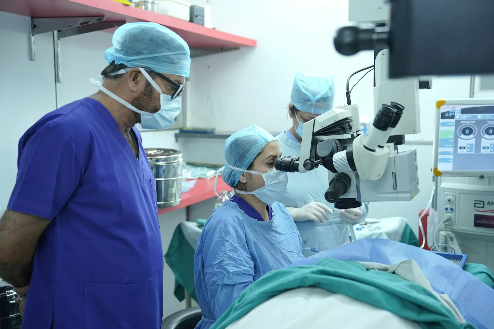
What are the steps of the procedure?
The procedure follows a precise, codified sequence. Each step prepares the next, from the micro-access to the eye through to the placement of the implant that will replace the removed lens.
The micro-incision
The surgeon makes an incision of 2 to 3 mm at the junction between the cornea and the white of the eye. This opening, described as self-sealing, generally closes without a suture. Its small size is one of the great advantages of phacoemulsification, because it limits deformation of the cornea and keeps surgically induced astigmatism to a minimum.
The capsulorhexis
The lens is contained within a thin, transparent envelope, the capsule. The surgeon cuts a regular circular opening in it: this is the capsulorhexis. This window gives access to the nucleus of the lens while preserving the capsular bag, which will later hold the implant. The precision of the capsulorhexis directly determines the stability of the implant’s positioning in the weeks that follow.
Ultrasound fragmentation and aspiration
The ultrasound probe is introduced through the micro-incision. It breaks up the nucleus of the lens, while an irrigation-aspiration system removes the debris as it goes. The capsular bag is left in place, like an empty setting ready to receive the implant. A viscoelastic — a protective gel-like substance — is injected before and after fragmentation to protect the delicate structures of the eye, notably the corneal endothelium.
Placement of the intraocular implant
A soft intraocular lens is finally injected, folded, through the same micro-incision. It unfolds within the capsular bag and settles into the place of the removed lens. The power of the implant is calculated beforehand during the pre-operative assessment (ocular biometry), in order to aim for the desired visual correction. Depending on the patient’s visual needs, several types of implant exist, detailed on our dedicated page on premium implants.
ANAESTHESIA
Anaesthesia and duration of the operation
Phacoemulsification is most often carried out under topical anaesthesia: simple anaesthetic eye drops are instilled into the eye, making the ocular surface insensitive without an injection. The patient remains awake, without feeling any painful gesture during the procedure. A peribulbar or retrobulbar injection (injectable local anaesthesia) may be preferred in certain situations. General anaesthesia remains reserved for particular cases that justify deep sedation.
The procedure itself is short: it generally lasts 15 to 30 minutes for one eye. It is performed as day-case surgery, which means the patient comes in and leaves the same day, without staying overnight at the clinic. The total time spent on site — preparation, procedure, monitoring — is longer than the operating time itself.
With Dr Tourabaly, this surgery is performed at the Clinique Sainte-Geneviève, in Paris 14. The two eyes are not operated on the same day. One eye is treated first, then the second after an interval of a few days to a few weeks, once the first eye is well into its recovery.
RECOVERY
What is recovery like?
Visual recovery is often quick, but it remains gradual and varies from one patient to another. In the first few days, vision may be blurred and the eye a little red or sensitive to light. These sensations usually fade over the days as the eye heals and the implant settles into its final position.
A course of anti-inflammatory and antibiotic eye drops is prescribed for several weeks to support healing and prevent infection. A few precautions are recommended: avoid rubbing the eye, do not expose the eye to uncontrolled water (swimming pool, sea) for about a month — until the anti-inflammatory drops have reduced local defences — and limit intense effort in the first few days.
Returning to driving depends on each patient’s own recovery and on the state of the other eye; it is assessed at the post-operative follow-up. There is no fixed interval applicable to everyone. The management of the post-operative course is detailed on the cataract surgery page.
Practical post-operative point
Like any intraocular surgery, phacoemulsification carries risks that are rare but real (infection, detachment, raised intraocular pressure). These risks are systematically explained in consultation before the procedure. The decision to operate is always taken with full awareness, after a personalised examination.
TECHNIQUES
Phacoemulsification and femtosecond laser
For some years now, a femtosecond laser has been able to assist certain steps of cataract surgery. This is referred to as femtosecond laser-assisted cataract surgery (FLACS, from the English acronym). The laser performs part of the gestures traditionally carried out by hand by the surgeon.
The laser can thus perform the incision, the capsulorhexis and a pre-fragmentation of the lens nucleus, with reproducible precision. The fine fragmentation and aspiration, however, remain carried out by the ultrasound of phacoemulsification. The two techniques are not in opposition: the laser is a complement, on specific steps.
The respective place of these two approaches is the subject of evaluations by the health authorities, notably the Haute Autorité de Santé (HAS). The choice of technique is discussed case by case, according to the characteristics of the eye, the type of cataract and each patient’s file, during the pre-operative consultation. Recent innovations in this field are presented on the technological innovations in eye surgery page.
Torsional phaco and other developments
The phacoemulsification equipment itself has undergone developments. The so-called torsional technique (Ozil, on Alcon machines) uses a rotational movement of the probe rather than a back-and-forth movement. This mode reduces the risk of damaging the cells of the corneal endothelium and lowers the total energy delivered into the eye. Other modes (burst, pulse) also make it possible to modulate energy delivery according to the hardness of the nucleus. These parameters are adjusted by the surgeon according to the specific characteristics of each lens.
INDICATIONS

When is phacoemulsification indicated?
Phacoemulsification is offered when the cataract sufficiently impairs vision and daily life: difficulty reading, glare, reduced night vision, dulled colours, halos around light sources. There is no universal threshold based on a visual acuity figure: the decision rests on the real impact assessed in consultation, cross-referenced with the patient’s expectations and lifestyle.
Before any procedure, a pre-operative assessment is carried out: a complete examination of the eye, measurements (biometry) to calculate the power of the implant, and a search for any anatomical particularities. This assessment also makes it possible to identify the symptoms of cataract and to know when to consult.
- Age-related cataract (the most common): progressive clouding of the lens from around the age of sixty, sometimes earlier.
- Cataract secondary to a disease: diabetes, prolonged corticosteroid treatment, chronic uveitis.
- Post-traumatic cataract: clouding occurring after an ocular injury.
- Congenital cataract: rarer, requiring specialist care from childhood.
To learn about the complete care pathway, the assessment and the consultation arrangements, see our dedicated page on cataract surgery. The decision to operate is always taken after a personalised examination.
FAQ
Frequently asked questions
Is phacoemulsification painful?
The procedure is carried out under topical anaesthesia — simple anaesthetic eye drops that make the ocular surface insensitive — so that the procedure is not felt as painful. The patient may perceive light and slight sensations of pressure during the procedure. After the operation, temporary discomfort is possible in the first few days, eased by the prescribed treatment.
How long does the procedure take?
Phacoemulsification generally lasts 15 to 30 minutes for one eye. It is performed as day-case surgery: the patient comes in and leaves the same day. The total time spent at the clinic is longer than the operating time, because of the preparation and the immediate post-operative monitoring.
Should both eyes be operated on the same day?
No. The two eyes are not operated on simultaneously. One eye is treated first, then the second after an interval of a few days to a few weeks, once the first eye is well into its recovery. This interval is tailored to each patient.
What is the difference between phacoemulsification and femtosecond laser?
The femtosecond laser can assist certain steps (incision, capsulorhexis, pre-fragmentation of the nucleus), but the fine fragmentation and aspiration of the lens remain carried out by the ultrasound of phacoemulsification. The two approaches are complementary, not opposed. The choice is discussed at the pre-operative assessment according to the characteristics of the eye.
Can the cataract come back after the operation?
The removed lens does not re-form. However, the capsule left in place may become opaque over time: this is what is known as secondary cataract. It is treated with a simple laser procedure in consultation (YAG laser), without further surgery in the operating theatre.
When can driving be resumed after phacoemulsification?
There is no fixed universal interval. The resumption of driving depends on the visual recovery of the operated eye and on the quality of vision of the other eye. It is assessed at the post-operative follow-up. In practice, many patients resume driving in the first few days after the procedure, once their visual acuity allows it.
Where does this surgery take place with Dr Tourabaly?
Cataract surgery by phacoemulsification is performed at the Clinique Sainte-Geneviève, in Paris 14, as a day case. The consultation, the pre-operative assessment and the post-operative follow-up are organised beforehand. The full details of the care pathway are set out on our dedicated page on cataract surgery.
Sources
- Société Française d’Ophtalmologie (SFO) — Cataract surgery, phacoemulsification and intraocular implants.
- Haute Autorité de Santé (HAS) — Evaluation of cataract surgery and femtosecond laser-assisted surgery.
- Collège des Ophtalmologistes Universitaires de France (COUF) — Crystalline lens, cataract and surgical management.
- Ameli.fr (Assurance Maladie) — Cataract: symptoms, treatment and care.
This article is intended for information and does not replace a medical consultation. Only an ophthalmological examination can assess your cataract, establish the surgical indication and provide a personalised diagnosis.
A question about your cataract?
Dr Moïse Tourabaly, a former chef de clinique at the Quinze-Vingts hospital, carries out a complete assessment of your vision and informs you about the solutions suited to your situation — at the Clinique Sainte-Geneviève, Paris 14.
Patient journey: your cataract surgery step by step
Written and reviewed by Dr Moïse Tourabaly, ophthalmic refractive surgeon — former chef de clinique (Quinze-Vingts National Eye Hospital).
Last updated: September 11, 2026