
Monitoring tool
Amsler grid: testing your central vision at home
A grid you look at with one eye at a time. If the lines seem wavy, blurred or broken, your macula deserves an examination.
- 1Set the screen
- 2Observe
- 3Result
Setting your screen to its real size
A screen does not know its own size. Hold a bank card (or any card of the same format) vertically against the screen, then drag the slider until the blue rectangle is exactly the size of the card.
No card to hand? Skip this step: the grid will use an average size, slightly less accurate.
Prefer it on paper? A grid 10 cm across, at actual size, to be viewed from 30 cm — one eye at a time, with your reading glasses.
Looking at the central dot, one eye at a time
- Keep your reading glasses on if you wear them.
- Hold the screen 30 cm from your eyes, in good light.
- Cover one eye with your palm, without pressing on it.
- Fix your gaze on the central dot and never look away from it.
- Without moving your gaze, spot the areas where the lines seem wavy, blurred, grey or missing, then tap them on the grid.
0 area(s) marked on this eye.
Is this change recent?
This is the most important question: a distortion that has appeared recently is not to be monitored at home, it is to be shown.
Result
What your test shows
No area marked on either eye
Your central vision shows no distortion today, on this test. That does not replace an examination: the Amsler grid sees neither the periphery of the retina, nor glaucoma, nor an early change that only OCT reveals.
If you are being followed for AMD, diabetic retinopathy or high myopia, repeat this test regularly, always under the same conditions.
Areas are marked, and you already know about them
A stable distortion, already known to your ophthalmologist, is monitored. Note the marked areas and how they change, and bring that information to your next consultation.
If the areas spread from one test to the next, or if a new one appears, do not wait for that appointment: get back in touch.
Areas are marked, and they seem recent
A distortion of the lines (metamorphopsia) or a missing patch (scotoma) that has appeared recently warrants a prompt ophthalmological opinion, without waiting for a scheduled appointment. Several macular conditions are treated all the better for being caught early.
If it appeared suddenly, within a few hours, or if your vision dropped abruptly, this is an emergency: contact an ophthalmological emergency service the same day.
In every case — a sudden drop in vision together with slurred speech, weakness in a limb or an unusual headache means calling 15, the French emergency medical number: it may be the equivalent of a stroke.
A 10 cm grid: view it from 30 cm, one eye at a time, fixing your gaze on the central dot, with your reading glasses.
This test requires JavaScript. You can print a standard Amsler grid and view it from 30 cm, one eye at a time, fixing your gaze on the central dot.
What the Amsler grid shows — and what it does not
The grid explores about 20° of the central visual field, that is to say the macula. It brings out two signs: metamorphopsia, when straight lines appear wavy or distorted, and scotoma, when an area appears grey, blurred or absent.
Its limits are real and worth knowing. The test is subjective: the brain spontaneously fills in missing areas, which can mask a scotoma (perceptual filling-in). It does not see the peripheral retina, so neither an early detachment nor a tear. It screens for neither glaucoma nor cataract. A normal test therefore never replaces a fundus examination or an OCT.
Who is this test useful for?
Amsler grid monitoring is offered mainly to people followed for AMD, particularly when one eye is already affected and the other must be watched. It is also useful in diabetic retinopathy with macular involvement, epiretinal membrane, high myopia, or after certain retinal procedures.
It has no screening value in the general population without a risk factor: carried out in isolation, it worries more than it informs. Its proper use is regular, comparative monitoring, under the same lighting and at the same distance, by someone whose ophthalmologist has recommended it.
How to interpret what you see
- The lines are straight and even, the grid is complete — a normal test today, on this eye.
- The lines wave, bend or draw closer together — metamorphopsia. This is the most characteristic sign of a macular condition.
- An area is grey, dark or empty — scotoma. The central part is the most significant.
- The squares look smaller or larger on one side — micropsia or macropsia, which also reflect macular suffering.
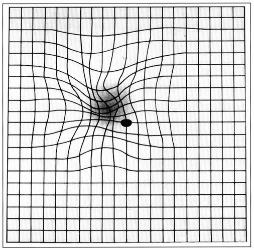
An abnormal result establishes no diagnosis. It indicates that an examination is called for, and how soon: promptly if the sign is new, the same day if it appeared suddenly.
Printable Amsler grid (PDF)
Do you prefer paper, to put it up on the refrigerator or keep it next to your reading chair? Download the printable Amsler grid (PDF, one A4 page). Print it at 100%, with no scaling: the whole grid must measure 10 cm on each side. The instructions for use are on the sheet.
Amsler grid and glaucoma: the right test?
No. The Amsler grid explores the center of the visual field, whereas glaucoma first affects the visual field outside the center and remains without symptoms for a long time. A normal grid test therefore says nothing about glaucoma. Glaucoma screening and monitoring rely on four examinations, all carried out during a consultation: measuring the pressure of the eye, examining the optic nerve, OCT and automated visual field testing.
Frequently asked questions about the Amsler grid
A new sign is to be shown, not monitored
Dr Moïse Tourabaly sees patients in consultation at the Cachan practice and at the Diabet’ Paris 13 practice for examination of the retina and the macula. A fundus examination and an OCT can state what the Amsler grid can only suggest.
Book an appointment on Doctolib — or browse the other tools and resources for understanding your sight.