Diplopia: when and why you see double
Diplopia is the perception of two images of a single object, in other words the experience of “seeing double”. It is a symptom, not a disease in itself. It can be harmless and caused by a simple optical problem within the eye, or it can reveal a neurological condition requiring urgent care. Telling these two situations apart is the starting point of any assessment.
UNDERSTANDING
What is diplopia?
Diplopia is the medical term for double vision: a single object is perceived as doubled, with two distinct images. These images may lie side by side (horizontal diplopia), one above the other (vertical diplopia), or slightly tilted (oblique diplopia). The offset may be subtle or very marked depending on the cause.
Normally, each eye sends the brain a slightly different image, and the brain merges them into a single three-dimensional perception. For this fusion to work, both eyes must be perfectly aligned and light must pass through the ocular media without being deflected. When this balance is disrupted — whether at the optical level or at the motor level — diplopia appears. Understanding at which level the disruption occurs guides the entire work-up.
Two broad types are distinguished, and telling them apart is crucial: monocular diplopia and binocular diplopia. This distinction takes just a few seconds with a simple test, and it determines how urgent the situation is.
KEY DISTINCTION
Monocular or binocular diplopia: the essential distinction
This is the most important distinction, because it separates the usually harmless causes from the potentially serious ones. The test is simple and immediate: the person is asked to cover one eye, then the other. Depending on whether the double vision persists or disappears, the diagnosis points in two radically different directions.
Monocular diplopia: an optical cause within the eye
Monocular diplopia persists when the other eye is closed: it remains present with a single eye open. It is almost always optical in origin, linked to an abnormality in the transparent media of the affected eye. By covering each eye in turn, you can identify the eye responsible: it is the one that, on its own, keeps seeing double. This form is most often harmless, but it still warrants an ophthalmological examination to find the cause and correct it.
Binocular diplopia: a misalignment of the eyes
Binocular diplopia disappears as soon as either eye is closed: it exists only with both eyes open. It reflects a misalignment of the two eyes, whose visual axes no longer point towards the same object. The brain then receives two images that cannot be overlaid and can no longer fuse them. It is this form which, particularly when it appears suddenly, can be a sign of damage to the oculomotor nerves or the central nervous system.
The one-eye test in practice
Cover one eye with your hand. If the double vision disappears → binocular diplopia (alignment at fault). If it persists with a single eye → monocular diplopia (an optical problem in the affected eye). This simple test determines how urgent the situation is: sudden binocular diplopia is a medical emergency until proven otherwise.
EMERGENCY

Sudden binocular diplopia: when to call emergency services?
EMERGENCY — Call the emergency services (dial 15 / the Samu in France) or go to A&E without delay if the double vision is accompanied by:
- An intense, sudden, unusually severe headache (a “thunderclap” headache)
- A sudden drooping eyelid (ptosis) or a pupil that is larger on one side
- Weakness on one side of the body, numbness, or difficulty with speech or balance
- Associated nausea, vomiting or reduced alertness
- Diplopia occurring after a head or facial injury
These associated signs may indicate a stroke, an aneurysm, or an oculomotor palsy of neurological origin. Every hour counts.
Apart from these warning signs, monocular diplopia that has come on gradually, or an isolated, intermittent nuisance, calls for a scheduled ophthalmological consultation. If you are unsure whether it is monocular or binocular, seek medical advice promptly rather than waiting.
CAUSES
What are the most common causes?
The causes fall into groups according to the type of diplopia. Monocular diplopia points to an optical problem in a single eye; binocular diplopia points to a misalignment of the two eyes, which may be muscular, nerve-related or neurological in origin. Here are the causes most often seen in practice.
Causes of monocular diplopia
These causes affect the path of light inside a single eye, between the cornea and the retina:
- Cataract: the progressive clouding of the lens can scatter light and double the image in a single eye. It is one of the most common causes of monocular diplopia, often associated with other symptoms such as glare and reduced contrast (see cataract surgery).
- High or irregular astigmatism: a cornea whose curvature is not perfectly spherical can split the image into two distinct points of focus. Uncorrected or poorly corrected astigmatism can thus cause subtle but bothersome doubling.
- Dry eye: an unstable tear film makes the corneal surface irregular, which can blur or double the image in a fluctuating way, particularly at the end of the day or in front of a screen (see dry eye).
- Keratoconus: this progressive deformation of the cornea creates a surface irregularity that multiplies the points where light converges. Monocular diplopia can be one of the first signs.
- An optical correction fault: unsuitable glasses or contact lenses, or a scratched or poorly centred lens, can cause a doubled image in one or both eyes.
Causes of binocular diplopia
These causes affect the alignment of the eyes, controlled by six oculomotor muscles per eye and by the cranial nerves that supply them:
- An oculomotor palsy: damage to the oculomotor nerve (III), the trochlear nerve (IV) or the abducens nerve (VI) prevents one or more muscles from working normally, misaligning the visual axes. These palsies may be of diabetic, vascular or neurological origin.
- Neurological causes: stroke, aneurysm, intracranial tumour, intracranial hypertension. It is these causes that make sudden binocular diplopia a neurological emergency.
- Systemic diseases: diabetes can cause oculomotor palsies (see diabetic retinopathy and diabetes); a thyroid disorder (thyroid eye disease) can infiltrate the muscles and restrict them; myasthenia, a neuromuscular disease, often produces fluctuating diplopia with ptosis.
- A long-standing oculomotor imbalance that decompensates: certain phorias (a slight misalignment usually compensated for by the brain) can decompensate abruptly with fatigue, illness or injury.
- Orbital trauma: a blow to the orbit can fracture the floor or mechanically trap a muscle, preventing the eye from moving normally.
INVESTIGATION
How is diplopia investigated?
The investigation begins with a precise history: how long it has been present, whether it came on suddenly or gradually, whether it is isolated or accompanied by other signs, constant or intermittent, and worse in certain directions of gaze. These answers strongly guide the diagnosis even before the clinical examination.
The first-line ophthalmological examination
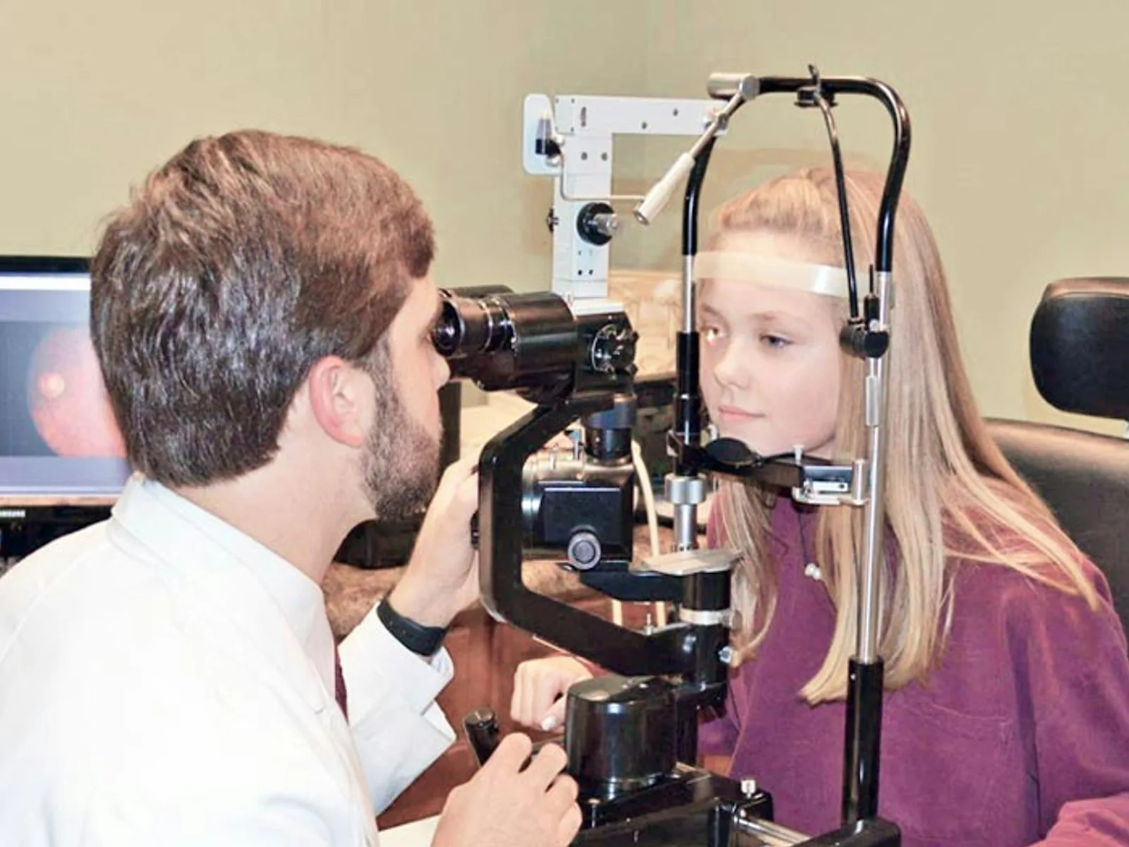
The ophthalmologist first carries out the fundamental test: covering each eye in turn to classify the diplopia as monocular or binocular. They then measure the refraction, examine the cornea and lens with the slit lamp, and study eye movements in the nine directions of gaze. For monocular diplopia, the examination focuses on the ocular media. For binocular diplopia, it assesses the field of gaze in which the diplopia is greatest, which helps identify the muscle or nerve involved.
The orthoptic assessment
The orthoptist completes the evaluation with precise measurements of the imbalance: angle of deviation for distance and near vision, the Lancaster test to map muscle deficits, and measurement of binocular fusion and stereopsis. This functional assessment is essential to quantify the disturbance, monitor its course and tailor management (prisms, rehabilitation).
Further investigations
Faced with binocular diplopia that appears neurological, brain imaging (MRI preferably, or an urgent CT scan) and a neurological opinion are requested promptly. Blood tests may look for diabetes, a thyroid disorder or myasthenia. If an associated acute ocular condition is suspected, ocular ultrasound and retinal imaging can complete the work-up.
TREATMENT
What management, according to the cause?
As diplopia is a symptom, its treatment depends entirely on the cause identified. There is no universal treatment for double vision: the cause is treated, and the diplopia usually improves as a result. Management can range from a simple change of glasses to neurosurgery, depending on the origin.
- Monocular diplopia due to cataract: when the clouding of the lens is advanced enough to justify surgery, cataract surgery usually resolves the diplopia at the same time as it improves overall visual acuity.
- Astigmatism or correction fault: a new, suitable optical prescription or surgical treatment of the astigmatism (LASIK, PRK, toric implant) corrects the cause.
- Dry eye: instilling artificial tears, optimising the environment and, if necessary, a topical anti-inflammatory treatment help to stabilise the tear film.
- Recent oculomotor palsy: spontaneous recovery is possible over several weeks to a few months, particularly for palsies of diabetic or vascular origin. In the meantime, a stick-on prism on a lens or temporary patching of one eye can eliminate the double vision in daily life.
- Persistent, stable oculomotor imbalance: once the situation has stabilised and if it is disabling, orthoptic rehabilitation, permanent prisms incorporated into the lenses, or surgery on the oculomotor muscles may be offered depending on the case.
- Neurological or systemic cause: management is that of the underlying disease, in a specialist setting — neurology, endocrinology or haematology depending on the aetiology.
A full ophthalmological assessment is the standard starting point. It makes it possible to link the double vision to its cause and to choose the appropriate solution, from a simple adjustment of glasses to urgent specialist care.
FREQUENTLY ASKED QUESTIONS
Learn more
- Amblyopia (lazy eye)
- The orthoptist
- The visual field test
- The anatomy of the eye
- Astigmatism
- Light halos
Frequently asked questions about diplopia
Is seeing double always serious?
No. Monocular diplopia, which persists with a single eye open, is most often optical in origin and harmless (cataract, astigmatism, dry eye). By contrast, sudden binocular diplopia — which disappears when one eye is closed and is accompanied by headache, ptosis or neurological signs — is a medical emergency.
How can I tell whether my diplopia is monocular or binocular?
Close one eye, then the other. If the double vision disappears as soon as one eye is closed, it is binocular and reflects a misalignment of the two eyes. If it persists with a single eye open, it is monocular and comes from an optical problem in that particular eye.
When is diplopia a neurological emergency?
Binocular diplopia of sudden onset is an emergency until proven otherwise, especially if it is accompanied by a severe headache, a drooping eyelid, an asymmetric pupil, weakness on one side of the body or difficulty with speech. These associated signs suggest a stroke, an aneurysm or an oculomotor palsy of neurological origin. Call the emergency services without delay.
Can a cataract cause double vision?
Yes. Clouding of the lens can scatter light and cause monocular diplopia, visible even with the other eye closed. It is a common cause. Treatment is by cataract surgery when the functional disturbance justifies it.
Can diplopia go away on its own?
It depends on the cause. Some recent oculomotor palsies — particularly those linked to diabetes or a vascular cause — recover spontaneously over several weeks to a few months. Others require active treatment of the cause. Regular ophthalmological follow-up allows the course to be assessed and management to be adjusted.
What can be done while the cause is being treated?
When binocular diplopia is troublesome in daily life but not urgent, temporary solutions can be offered: a stick-on prism placed on a spectacle lens or covering one eye with an opaque patch. These measures comfortably eliminate the double vision while the cause is being treated or the situation stabilises.
Can a child have diplopia?
Yes, but a child rarely complains spontaneously of seeing double: they often suppress one of the two images unconsciously, which can lead to amblyopia (reduced acuity in the suppressed eye). Any sign of squint or unusual visual behaviour in a child warrants an early ophthalmological consultation.
Sources
- French Society of Ophthalmology (SFO) — Diplopia, ocular motility and oculomotor palsies.
- College of University Ophthalmologists of France (COUF) — Semiology of diplopia, oculomotor and orthoptic examination.
- French National Authority for Health (HAS) — Identifying neurological warning signs and the emergency care pathway.
- Ameli.fr (French Health Insurance) — When to seek emergency care for a visual disturbance.
This article is for information purposes and does not replace a medical consultation. Binocular diplopia of sudden onset, especially when accompanied by severe headache, a drooping eyelid, pupil asymmetry or neurological signs, is an emergency: call the emergency services (dial 15 / the Samu in France) or go to A&E immediately. Only a medical examination can establish a personalised diagnosis.
Double vision that worries you?
Dr Moïse Tourabaly, ophthalmologist and former chef de clinique, carries out a full examination of your vision and directs you to the appropriate care. In the event of sudden diplopia with associated signs, call the emergency services first (dial 15 / the Samu in France).
Written and reviewed by Dr Moïse Tourabaly, ophthalmic refractive surgeon — former chef de clinique (Quinze-Vingts National Eye Hospital).
Last updated: September 1, 2026