Keratitis: causes, symptoms and treatment of corneal inflammation
Keratitis is inflammation of the cornea, the clear lens at the front of the eye. It can be mild or, conversely, sight-threatening depending on its cause. A red, painful, light-sensitive and watering eye should prompt prompt ophthalmological advice, particularly in contact-lens wearers, in whom certain infectious forms of keratitis progress rapidly.
UNDERSTANDING
What is keratitis?
Keratitis refers to any inflammation of the cornea, the transparent membrane at the front of the eye, in front of the iris and the pupil. The cornea plays a major optical role: it is through it that light enters the eye. Its transparency and regularity are essential for clear vision.
The cornea is also one of the most richly innervated tissues in the body. This explains why keratitis is often painful and accompanied by intense discomfort in the light. Its surface, the epithelium, forms a protective barrier against micro-organisms. When this barrier is weakened, an infection can take hold.
Two broad families of keratitis are distinguished, and their management differs radically:
- Infectious keratitis: caused by a micro-organism (bacterium, virus, fungus, amoeba). It can be serious.
- Non-infectious keratitis: linked to severe dryness, UV exposure, a foreign body or mechanical injury.
Depending on the depth of the injury, keratitis is described as superficial, limited to the epithelium, or deep (stromal), which affects the inner layers and carries a greater risk of scarring. Only an ophthalmological examination can determine the type and severity.
SYMPTOMS
What are the symptoms of keratitis?
Keratitis typically presents as a red and painful eye, associated with marked discomfort in the light and watering. These symptoms reflect the irritation of a highly innervated cornea. Their onset, especially when rapid and one-sided, warrants an ophthalmological examination without delay.
The most common signs
The symptoms of keratitis often overlap, whatever the cause:
- Eye pain, sometimes sharp, described as a burning or stinging sensation
- Red eye, particularly around the cornea
- Photophobia: discomfort in the light, or even an inability to tolerate it
- Profuse watering
- Foreign-body sensation, “like a grain of sand” under the eyelid
- Reduced vision or blurred vision when the central area of the cornea is affected
- Blepharospasm: difficulty keeping the eye open
The warning signs
Certain features point to a potentially serious form of keratitis. A whitish or greyish spot visible on the cornea may correspond to a corneal abscess. Purulent discharge, intense pain, a marked drop in vision or the rapid worsening of symptoms are all alarm signals.
In contact-lens wearers, any red and painful eye should be regarded as keratitis until proven otherwise. The lenses must then be removed immediately and urgent advice sought, without waiting for the situation to “sort itself out”.
A red, painful eye in a contact-lens wearer = an emergency — never put your lenses back in, do not mask the pain with an eye drop found in the medicine cabinet, and seek advice promptly. Infectious keratitis treated in time responds well; neglected, it can leave a scar on the cornea.
CAUSES
What are the causes of keratitis?
The causes of keratitis fall into two broad groups: infectious and non-infectious. The infectious forms, especially those linked to contact-lens wear, are among the emergencies in ophthalmology. Identifying the cause is decisive, because the treatment depends entirely on it.
Infectious keratitis
A micro-organism takes advantage of a break in the corneal epithelium to invade the cornea. Depending on the agent involved:
- Bacterial keratitis: the most common of the serious forms. Often linked to contact-lens wear, trauma or severe dryness. It can progress to a corneal ulcer.
- Viral keratitis, herpetic in particular: the herpes virus (HSV) causes a typical form of keratitis, known as dendritic, which can recur and leave lasting damage. Ophthalmic shingles is another viral cause.
- Amoebic keratitis: caused by an amoeba (Acanthamoeba) present in water. It is closely associated with contact-lens wear, especially where there is contact with tap, swimming-pool or shower water.
- Fungal keratitis: rarer, often after an injury caused by plant material (a branch, a thorn).
Non-infectious keratitis
The cornea can also become inflamed without infection, following a physical injury or an imbalance of its surface:
- Severe dry eye: an inadequate tear film weakens the epithelium and causes what is called “superficial punctate” keratitis. Prolonged screen work makes it worse (see our article on screen-related dry eye).
- UV exposure: the well-known “snow blindness”, after a day in the mountains without protection, or after exposure to a welding arc. UV rays burn the surface of the cornea.
- Foreign body: dust, metal filings or a grain of sand that abrades the cornea.
- Poor lens hygiene or prolonged wear, which mechanically irritate the cornea and expose it to infection.
- Eyelid abnormality or chronic blepharitis, which disrupt the tear film (see our article on blepharitis).
RISK FACTOR
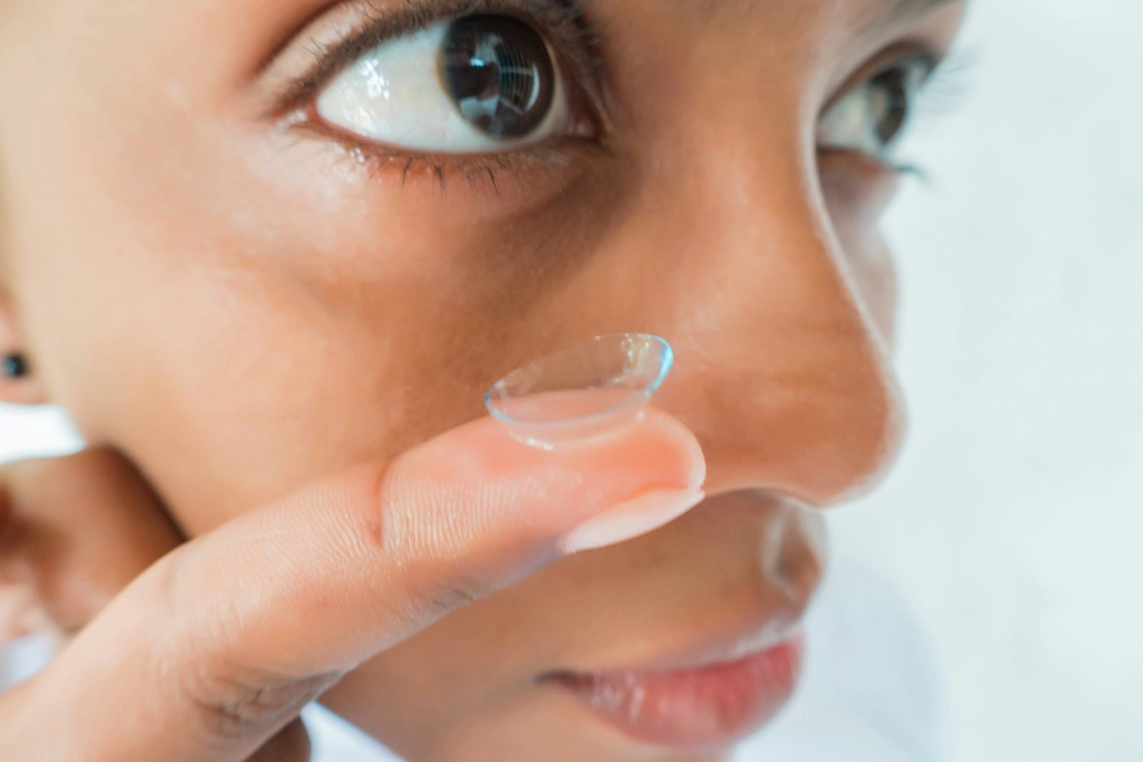
Why is contact-lens wear a major risk factor?
Contact-lens wear is the leading risk factor for infectious keratitis in young adults. The lens, placed directly on the cornea, can create micro-lesions, reduce oxygenation and act as a support for micro-organisms. This is why hygiene and wearing rules are essential.
How the lens promotes infection
Several mechanisms combine. The lens can cause fine epithelial erosions, which open the door to bacteria. It reduces the supply of oxygen to the cornea, especially with prolonged or overnight wear. Finally, a poorly maintained lens and case become a reservoir of germs.
Contact with water is particularly dangerous. Showering, bathing or swimming with your lenses, or rinsing the case with tap water, exposes you to the risk of amoebic keratitis, an infection that is dreaded because it is difficult to treat. Water, even when drinkable, is not sterile.
The highest-risk behaviours
Certain habits multiply the risk of keratitis in contact-lens wearers:
- Sleeping in your lenses when they are not designed for it
- Exceeding the recommended wearing time (late replacement)
- Rinsing or storing lenses with water or saliva instead of the appropriate solution
- Bathing, swimming or showering with your lenses
- Handling your lenses without washing your hands
- Reusing storage solution from one day to the next
The lens wearer’s golden rule — never let water touch the lenses, never wear them while swimming or sleeping unless explicitly indicated, and remove them immediately at the first sign of a red or painful eye. If in doubt, take the lens out and seek advice: this is the approach that best protects the cornea.
EMERGENCY
Is keratitis an emergency?
Keratitis can be an emergency, particularly when it is infectious. As the cornea is essential to vision, an untreated infection can progress to a corneal abscess and leave a scar that permanently impairs sight. The outcome depends largely on how quickly it is managed.
The risk of corneal scarring
When the inflammation reaches the deep layers of the cornea, healing can leave an opacity. If this opacity lies within the visual axis, at the centre of the cornea, it causes lasting interference with vision. This is precisely what is at stake with early treatment: preserving the transparency of the cornea.
In the most severe forms, infectious keratitis can be complicated by a corneal perforation or by spread of the infection inside the eye. These situations, fortunately rare, underline the importance of never dismissing a red and painful eye that is getting worse.
When to seek advice without delay
Seek advice promptly, as an emergency, if any of these signs are present:
- A red and painful eye in a contact-lens wearer
- A white or grey spot visible on the cornea
- Reduced vision associated with the pain
- Intense photophobia preventing the eye from opening
- Purulent discharge or rapid worsening of symptoms
DIAGNOSIS
How is keratitis diagnosed?
The diagnosis of keratitis rests on the slit-lamp examination, often supplemented by a fluorescein test. This examination, which is not painful, allows the ophthalmologist to observe the cornea in detail, to measure the extent of the injury and to point towards the cause. It is the key step in management.
The slit-lamp examination
The slit lamp is a microscope that projects a thin beam of light onto the eye. It makes it possible to examine the cornea layer by layer, to detect an erosion, an infiltrate or an abscess, and to assess the state of the anterior chamber. It is the reference tool for evaluating keratitis.
The fluorescein test
A drop of yellow-orange dye, fluorescein, is instilled into the eye. Under a blue light, it highlights the areas where the corneal epithelium is damaged, which then appear coloured. This test reveals the characteristic branching (dendritic) pattern of herpetic keratitis or the extent of an erosion.
Where a severe infection is suspected, particularly with an abscess, a corneal scraping may be taken to identify the germ in the laboratory and adjust the treatment. This step is decided by the ophthalmologist according to the clinical presentation.
TREATMENT
How is keratitis treated?
The treatment of keratitis depends entirely on its cause. Bacterial keratitis calls for antibiotic eye drops, herpetic keratitis for antivirals, dry keratitis for artificial tears. There is therefore no single treatment: it is the ophthalmological diagnosis that guides the prescription, never self-medication.
Treatment tailored to the cause
Depending on the origin identified, the ophthalmologist may prescribe:
- Antibiotic eye drops for bacterial keratitis, sometimes to be instilled very frequently at the start
- Antiviral treatment (drops or tablets) for herpetic keratitis
- Antifungals for fungal keratitis, or specific prolonged treatment for amoebic keratitis
- Artificial tears and treatment of dryness for dry keratitis
- Stopping lens wear for the whole duration of treatment and until healing is complete
The danger of self-medication
One point deserves particular attention: never self-medicate with a cortisone eye drop. Corticosteroids can considerably worsen infectious keratitis, herpetic keratitis in particular, and promote an abscess. An eye drop that works on one red eye can be dangerous on another.
Likewise, the pain should not be “masked” with a local anaesthetic, nor should lenses be put back in to see clearly. Any drop instilled into an inflamed eye must be given on prescription, after examination. This is the fundamental safety rule when faced with keratitis.
Prevention in contact-lens wearers
Prevention remains the first treatment. In contact-lens wearers, a few simple rules markedly reduce the risk of keratitis:
- Wash and dry your hands before any handling
- Respect the wearing and replacement schedule
- Use only the appropriate cleaning solution, never water or saliva
- Renew the case regularly and let it air-dry
- Do not sleep, swim or shower with your lenses
- Have a regular ophthalmological check of the lens fit
FAQ
On the same topic
Frequently asked questions
Is keratitis serious?
It depends on the cause. Keratitis linked to dryness or UV is often mild and heals quickly. Infectious keratitis, particularly in contact-lens wearers, can be serious and threaten vision if it is not treated promptly. Only an ophthalmological examination can settle the matter.
How long does keratitis last?
Mild superficial keratitis can heal within a few days with appropriate treatment. Deep infectious keratitis requires longer management and close monitoring. The duration depends on the cause and the depth of the injury, which is why early advice is valuable.
Can you wear your lenses with keratitis?
No. Lens wear must be stopped immediately as soon as symptoms appear and throughout the whole treatment, until healing is complete and confirmed by the ophthalmologist. Putting your lenses back in too soon risks worsening and recurrence.
Which eye drop should be used for keratitis?
There is no single eye drop: the treatment depends on the cause (antibiotic, antiviral, antifungal, artificial tears). Above all, never use a cortisone eye drop without advice, as it can worsen infectious keratitis. Every eye drop should be prescribed after an ophthalmological examination.
Is keratitis contagious?
Keratitis itself is not contagious from one eye to another by simple contact. However, some causes, such as the herpes virus, can be transmitted. Good hand hygiene and not rubbing the eye remain useful precautions.
Why are contact-lens wearers more at risk?
The lens can create micro-lesions of the cornea, reduce its oxygenation and harbour germs if it is poorly maintained. Contact with water additionally exposes wearers to amoebic keratitis. Strict adherence to hygiene rules markedly reduces this risk.
Is diagnosing keratitis painful?
No. The slit-lamp examination and the fluorescein test are not painful. The dye may give a slight, passing sensation of coolness. This examination allows the cornea to be analysed in detail and the treatment to be directed at the Paris 13 or Cachan practice.
Scientific sources
- French Society of Ophthalmology (SFO). Infectious keratitis and corneal disease. SFO report and recommendations.
- College of University Ophthalmologists of France (COUF). Ophthalmology — Item: red and painful eye, corneal disease. National reference syllabus.
- French National Authority for Health (HAS). Management of the red eye and of keratitis: good-practice recommendations.
- French National Health Insurance (Ameli.fr). Keratitis and contact-lens wear: symptoms, prevention and what to do. Information sheet.
- Inserm. Cornea and ocular surface: information dossier.
Further reading
- Dry eye and screens: understanding and relieving it
- Keratoconus: recognising the early signs in young adults
- Blepharitis: inflammation of the eyelids and the tear film
- Book an appointment at the practice
Cachan practice · Tel. +33 1 45 47 08 11
Paris 13 practice · Tel. +33 1 89 31 30 60
Medical disclaimer
This article is for information purposes. Personalised ophthalmological advice remains essential for any therapeutic decision.
This article is intended for general information and does not replace a medical consultation. Diagnosing keratitis and identifying its cause require a complete ophthalmological examination, with a slit lamp and a fluorescein test. Faced with a red and painful eye, discomfort in the light or reduced vision, particularly in a contact-lens wearer, remove your lenses and seek advice promptly from an ophthalmologist. Never take a cortisone eye drop without medical advice.
—
Written and reviewed by Dr Moïse Tourabaly, ophthalmic refractive surgeon — former chef de clinique (Quinze-Vingts National Eye Hospital).
Last updated: September 5, 2026