Retinal transplant: a blind woman regains her sight, and what this world first really changes
In Turin, a surgical team transferred the macula and the anterior segment of one eye to the other eye of the same patient, who recovered partial vision after more than five years of blindness. The key word is not “retina”: it is “the same patient”. This was not a graft from a donor, but an autograft — and that distinction, which disappears from most press coverage, explains both why the operation could work and why it does not apply to the most common retinal diseases, age-related macular degeneration (AMD) chief among them.
WHAT HAPPENED
What the Turin team actually did
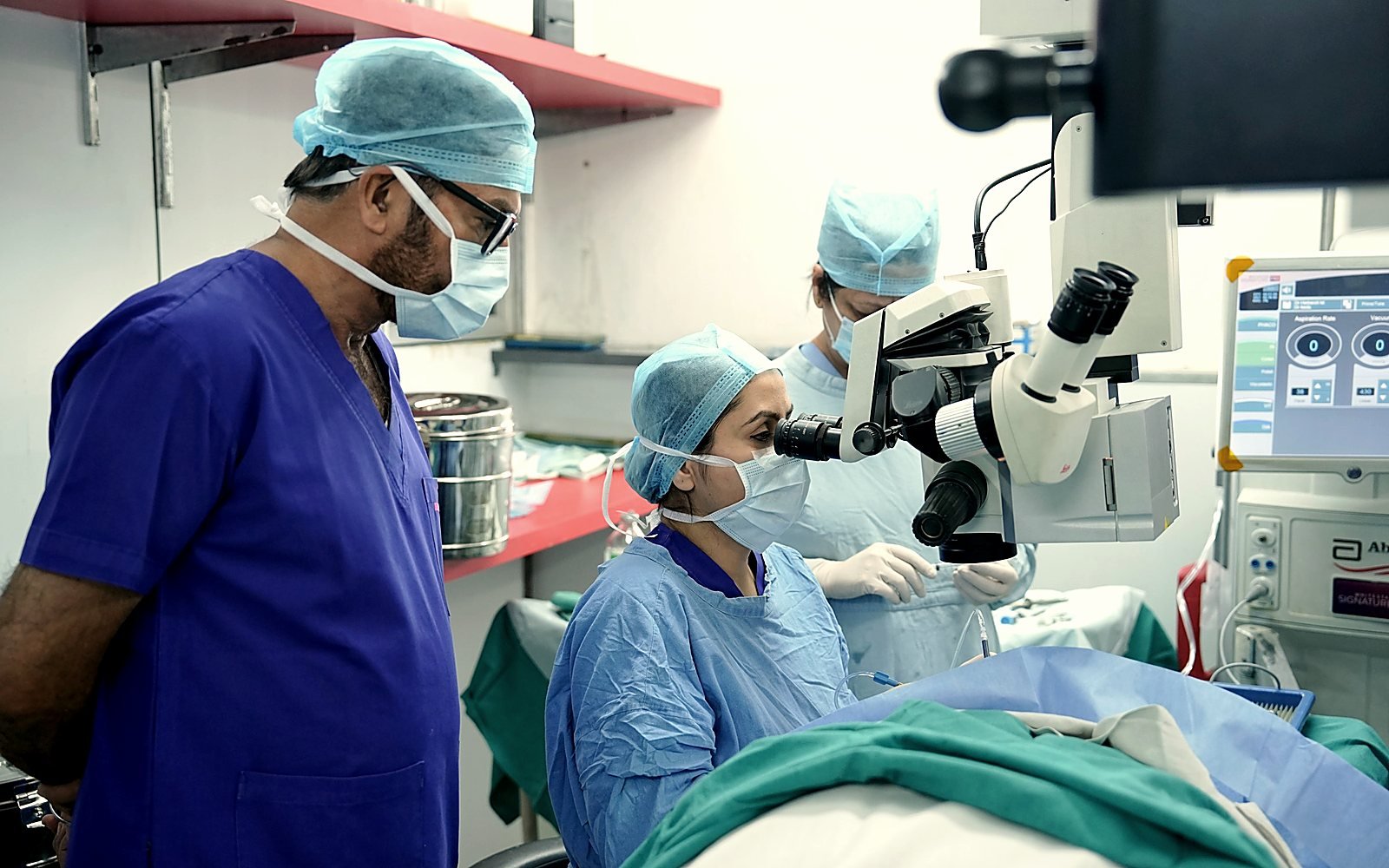
The Molinette hospital, part of the Azienda Ospedaliero-Universitaria Città della Salute e della Scienza in Turin, announced the operation on 26 July 2026. The surgery itself had been performed about three weeks earlier: the announcement was held back until the team could confirm that the graft was holding and that the patient really was recovering vision. The team was led by Professor Michele Reibaldi, head of the hospital’s university eye clinic. The patient, a 67-year-old woman, had been blind for more than five years.
A point of vocabulary needs making straight away, because headlines have circulated widely under the phrase “retinal transplant”. What was transplanted is not the whole retina, but an anatomical block comprising the cornea, the sclera, the conjunctiva and the central portion of the retina, that is, the macula. Put another way: the optical window at the front of the eye and the area of fine vision at the back travelled together, from one eye to the other, in a single procedure.
One patient, two eyes, two opposite problems
To understand why this operation makes sense, picture the eye as a chain of three links: an optical system at the front (cornea, lens) that forms the image, a sensor at the back (the retina, whose macula is the high-definition centre), and a cable (the optic nerve) that carries the signal to the brain. If a single link is broken, vision stops, whatever the state of the other two.
In this patient, both eyes were out of service, but for opposite reasons:
- The left eye had an irreparably damaged optic nerve following a road accident. The cable was cut. But the optics and the sensor had remained healthy: a clear cornea, a working macula, a perfectly formed eye that no longer served any purpose.
- The right eye, by contrast, had kept an optic nerve still able to conduct the signal, but suffered from a maculopathy made worse by injuries that had also compromised the transparency of the cornea. The cable was working; the optics and the sensor were destroyed.
The logic of the operation then becomes plain: take the intact links from the left eye and graft them onto the only cable still working, that of the right eye. It is this reasoning, more than the technical feat, that makes the case original.
A block of tissue, not a membrane
The surgical difficulty lies in the nature of what is being moved. A corneal graft handles a transparent lamella a few hundred microns thick, avascular, nourished by imbibition. Here, the transferred block contained vascularised tissue and a fragment of nervous tissue, the macula, whose survival depends on its blood supply. According to the technical descriptions published in the specialist press, the team is said to have preserved the long posterior ciliary arteries in order to maintain perfusion of the graft during the transfer; that detail appears in a single source, however, and has not been confirmed by the hospital’s press release.
The operation is reported to have lasted about six hours, a figure repeated by French-language news agencies but absent from the original Italian communications.
THE WORD THAT CHANGES EVERYTHING
Autograft or donor graft: why the difference is decisive
In medical language, an autograft is tissue taken from a patient and re-implanted in that same patient. An allograft is tissue that comes from another individual, most often a deceased donor. The whole of transplant medicine is organised around this distinction, because it determines how the immune system reacts.
A graft from a donor carries surface markers that the recipient’s body does not recognise as its own. It attacks them. This is rejection, which in most organ transplants requires lifelong immunosuppressive treatment, with everything that comes with it: increased risk of infection, regular blood monitoring, side effects. In the Turin case, that question simply does not arise. The tissue comes from the patient herself: her immune system has no reason to turn against it.
What this avoids in practical terms
This is a point I can illustrate from my daily practice of corneal grafting. The cornea is an immunologically privileged tissue: it contains no blood vessels, which partly shields it from immune surveillance. That is why a keratoplasty generally requires no systemic immunosuppression, where a kidney or liver transplant does. Despite that advantage, rejection remains the complication we fear and monitor for years, with prolonged corticosteroid eye drops and close follow-up.
Transpose that constraint to a graft containing retinal nervous tissue and vascularised tissue, neither of which enjoys any immune privilege: rejection would become the central obstacle. By setting that question aside, the Turin team removed one of the most unpredictable variables in the equation. It takes nothing away from the difficulty of the procedure, but it reframes what the case demonstrates — and what it does not.
CORNEA
Why a conventional corneal graft could not have been enough
One question arises naturally: if the cornea of the right eye was opaque, why not simply graft a donor cornea, as is done routinely? The answer comes in two parts.
The first is obvious: a new cornea would have been of no use as long as the macula behind it remained destroyed. Making the window clear does not restore vision if the sensor is out of service. The eye would have been cleared without its sight being restored.
The second is more technical and deserves explaining, because it also concerns patients I see in consultation. Corneal transparency is not a state acquired once and for all: it is maintained continuously by a population of stem cells housed in the limbus, the ring-shaped transition zone between the cornea and the white of the eye. These cells renew the corneal epithelium throughout life and act as a barrier: they stop the conjunctiva, which is opaque and vascularised, from invading the optical surface.
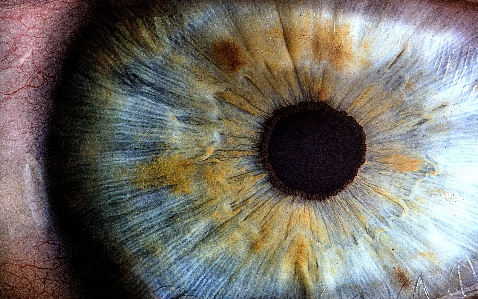
When that reservoir is destroyed — by a chemical burn, a severe injury or certain inflammatory diseases — the condition is called limbal stem cell deficiency. The cornea loses its capacity to repair itself, becomes gradually covered with opaque, vascularised conjunctival tissue, and turns into hostile ground. Placing a donor graft on it amounts to laying new tissue on soil that cannot sustain it: the graft becomes opaque in turn, and the failure rate is high.
It is precisely in these situations that surgery has turned, over the past twenty years or so, to autologous limbal stem cell grafts — taken, therefore, from the patient’s own healthy eye. A systematic review published in the British Journal of Ophthalmology reports for these techniques anatomical success of around 69% and functional success close to 60%, with a median follow-up of about one year and nine months.
Seen from that angle, the Turin operation follows an already familiar line of reasoning — going to the patient’s own body for the tissue that cannot be replaced by a graft from someone else — but pushes it far further, as far as taking the anterior segment and the macula in a single block.
SETTING THE RECORD STRAIGHT
No, retinal transplantation will not become a treatment for AMD
This is the question this news will raise in consultations, and it is better answered plainly: this technique cannot be transposed to age-related macular degeneration, nor to diabetic retinopathy, nor to most inherited retinal diseases. It is not a matter of resources or of development time. It is an impossibility in principle, and it rests on three conditions that were all met in this patient.
It takes a “donor” eye that is healthy but permanently unusable
This is the most restrictive condition. Removing the macula and the cornea from an eye means sacrificing it. The procedure is only conceivable if that eye can no longer see in any case, for a reason unrelated to the tissue being taken — here, an optic nerve destroyed by trauma. In someone with AMD or diabetic retinopathy, the disease affects both eyes, often fairly symmetrically. There is no expendable eye, and no healthy macula to be taken from anywhere.
It takes a working optic nerve in the recipient eye
The retina is not a tissue like any other: it is an extension of the central nervous system, and the optic nerve is a bundle of about a million nerve fibres. We do not know, today, how to reconnect those fibres surgically. A retinal graft can therefore transmit nothing at all unless it is placed in an eye whose optic nerve and cerebral visual pathways have remained able to conduct.
The clearest illustration of this limit comes from elsewhere. In 2024, the NYU Langone team published in JAMA the follow-up of a combined face and whole-eye transplant — a surgical feat without equal, with an eyeball that was revascularised and survived. The visual outcome, one year on, is summed up in a single sentence of the paper: no light perception in the transplanted eye. The globe was alive; the optic nerve had not resumed its function. Until that lock is opened, eye transplantation in the sense the public imagines will remain out of reach.
It takes a markedly asymmetrical condition
The two preceding conditions have to coexist in the same person, and as mirror images: the donating eye must have everything except the nerve, the receiving eye must have nothing but the nerve. That configuration is a matter of traumatic coincidence. It does not define a population of patients, but isolated cases. This is why the announcement should be read for what it is — the demonstration that a procedure considered impossible can be carried out — and not as the announcement of a treatment to come for common macular diseases.
PRECEDENTS
Moving a macula: an idea thirty years old
What is unprecedented in the Turin procedure is the transfer from one eye to the other. But the idea of moving the macula in order to take it out of a diseased area is an old one, and it went through genuine clinical development.
In the 1990s, before intravitreal injections arrived, macular translocation was developed to treat exudative AMD. The principle: detach the retina entirely, rotate it around the optic nerve, then lay it back down so that the macula rests on an area of pigment epithelium that is still healthy, away from the neovascular lesion. A series published in 1999 by Eckardt and colleagues in Graefe’s Archive reported that, of thirty eyes operated on, about 60% reached an acuity compatible with reading.
This technique is no longer performed today, and the reasons it was abandoned are instructive. A 2008 Cochrane review concluded that the evidence from randomised trials was insufficient, in a setting where complications were far from marginal: retinal detachment in six patients out of twenty-five in one of the series analysed, double vision requiring correction in five out of twenty-five.
Rotating the retina also rotates the image, which often calls for a second operation on the eye muscles. Then anti-VEGF treatments arrived, and major surgery gave way to an injection lasting a few minutes. The lesson still holds: a spectacular surgical innovation is not necessarily the one that will prevail.
CAUTION
What is still unknown about this case
We have to be clear about the status of this information. To date, this case has not been the subject of any publication in a peer-reviewed journal. Everything we know about it comes from the hospital’s press release and from its pick-up by the news agencies. This is not a criticism aimed at the team — the interval between an operation and its scientific publication is counted in months — but it does call for restraint.
Professor Michel Paques, head of the ophthalmology department at the Quinze-Vingts National Ophthalmology Hospital in Paris, confirmed to AFP that this was indeed a world first, while stressing that, in the absence of published and peer-reviewed data, it is not possible to assess objectively the technique, its reproducibility or the results announced. That is the right position, and it is the one I take here — all the more so as I practised in that hospital as Former Chef de Clinique at the Quinze-Vingts National Ophthalmology Hospital (Sorbonne University).
In concrete terms, several things are missing before any judgement can be made:
- No measured visual acuity has been released by the hospital. The reported outcome is descriptive: vision first in black and white, then in colour; the patient recognised her son’s face and her guide dog, and identified a banknote. An acuity figure is circulating on several news sites, but it appears in no primary source, so I will not repeat it.
- Follow-up is only a few weeks long. Yet the decisive questions are those of the medium term: will the grafted macula remain properly perfused? Will the corneal surface stay clear? Will visual function improve or deteriorate?
- Neither imaging nor a detailed protocol has been released — no OCT, no visual field, no full operative description that would let other teams assess how reproducible the procedure is.
Professor Reibaldi has himself stated that his patient would never regain fully normal vision. That reservation, coming from the surgeon, is probably the most useful sentence in the whole press pack.
IN PRACTICE
What actually exists today to preserve your sight
An announcement like this one often has a paradoxical effect: it feeds hope of a future treatment, and draws attention away from what is available now. Yet in the great majority of retinal diseases, most of the prognosis is decided by how early care begins, not by an exceptional technique.
- Exudative AMD: intravitreal anti-VEGF injections stabilise the disease and preserve acuity in a large proportion of patients, provided they are started early. Recent distortion of straight lines warrants a prompt opinion.
- Diabetic retinopathy: regular retinal screening in anyone with diabetes makes it possible to act before vision declines, that is, at a stage when the patient still feels nothing.
- Retinal detachment: the sudden appearance of floaters, flashes of light or a curtain in the periphery is an emergency. How quickly it is treated directly determines the visual outcome, particularly if the macula has not yet lifted.
- Epiretinal membrane: surgery that is properly indicated, and carried out before distortion becomes too established, gives a lasting improvement in visual comfort.
- Corneal opacities: modern lamellar grafts, DMEK in particular, replace only the failing layer of the cornea. They offer faster visual recovery and a lower risk of rejection than the full-thickness graft of the past.
None of this makes the front page. Yet this is where the difference between sight preserved and sight lost is decided, for the vast majority of patients.
FAQ
Frequently asked questions
Can a retina be transplanted to treat AMD?
No, and it is not a question of time. AMD affects both eyes: there is therefore no healthy macula to be taken from the same patient. The Turin operation was only possible because one eye had kept intact structures while being permanently blind through destruction of its optic nerve — a configuration that belongs to traumatic sequelae, not to a retinal disease. For AMD, management today rests on intravitreal injections in the exudative form and on monitoring in the atrophic form.
Is this really a retinal transplant?
Partly. The transferred block comprised the cornea, the sclera, the conjunctiva and the macula, that is, the central part of the retina — not the whole retina. The most accurate term is a combined autograft of the anterior segment and the macula. The phrase “retinal transplant” used by the press is a simplification that suggests a complete replacement of the visual sensor, which is not what happened.
Did the tissue come from a deceased donor?
No. That is the central point of the story: the tissue came from the patient’s own left eye, transferred to her right eye. This is called an autograft. That origin rules out the risk of immune rejection and avoids any immunosuppressive treatment, unlike a graft from a donor.
Will this technique be offered in France?
Nothing allows that to be said today. The case has not yet been published in a peer-reviewed journal, no imaging data and no detailed operative protocol have been released, and follow-up is counted in weeks. Before a technique enters practice, it has to be described, reproduced by other teams and assessed over time. Even supposing those steps are taken, the number of patients with the required anatomical configuration would remain very small in any case.
Why can a whole eye not simply be transplanted?
Because the obstacle is not the eye, but the optic nerve. That nerve is a bundle of about a million nerve fibres arising from the central nervous system, and we do not know how to reconnect them surgically. An American team performed a combined face and whole-eye transplant: the transplanted globe stayed vascularised and alive, but one year after the operation no light perception was found in that eye. That follow-up was published in JAMA in 2024. What is missing is nerve reconnection, not surgery.
What is the macular translocation that has been discussed since the 1990s?
It is a form of surgery developed before the era of intravitreal injections, which involved detaching the retina and then rotating it so as to reposition the macula over an area of pigment epithelium that was still healthy. It produced visual results in some patients, but at the cost of frequent complications — retinal detachment, double vision calling for additional muscle surgery. The arrival of anti-VEGF drugs, more effective and far less invasive, led to it being abandoned.
My central vision is declining: what should I do?
See an ophthalmologist without waiting for the outcome of ongoing research. A decline in central vision, distortion of straight lines or the appearance of a spot in the visual field warrants an eye examination with macular OCT, which identifies the origin of the problem. In most retinal diseases, the prognosis depends directly on how early the diagnosis is made — that is where the real leverage lies, far more than in exceptional techniques.
Scientific sources
- A.O.U. Città della Salute e della Scienza di Torino. Press release — ocular autograft performed at the Molinette hospital, 26 July 2026.
- Eckardt C, Eckardt U, Conrad HG. Macular rotation with and without counter-rotation of the globe in patients with age-related macular degeneration. Graefes Arch Clin Exp Ophthalmol. 1999;237(4):313-325. PMID: 10208265
- Eandi CM, Giansanti F, Virgili G. Macular translocation for neovascular age-related macular degeneration. Cochrane Database Syst Rev. 2008;(4):CD006928. PMID: 18843739
- Ziemssen F, Gelisken F. Macular translocation — a therapeutic approach for neovascular macular degeneration in the era of anti-VEGF therapy? Klin Monbl Augenheilkd. 2009;226(1):31-37. PMID: 19173161
- Shanbhag SS, Nikpoor N, Rao Donthineni P, Singh V, Chodosh J, Basu S. Autologous limbal stem cell transplantation: a systematic review of clinical outcomes with different surgical techniques. Br J Ophthalmol. 2020;104(2):247-253. PMID: 31118185
- Ceradini DJ, Tran DL, Dedania VS, et al. Combined Whole Eye and Face Transplant: Microsurgical Strategy and 1-Year Clinical Course. JAMA. 2024;332(18):1551-1558. PMID: 39250113
Further reading
- Dry AMD: OCT monitoring and practical advice
- Retinal detachment: recognising the emergency and acting fast
- Dr Moïse Tourabaly — Ophthalmologist
Cachan practice · Tel. +33 1 45 47 08 11
Medical disclaimer
This article is intended for information only. A personalised ophthalmological opinion remains essential for any treatment decision.
This article is intended as general information and does not replace a medical consultation. It describes an isolated clinical case, announced by press release and not yet published in a peer-reviewed journal; it therefore does not describe a treatment option that is available. Any decline in vision, distortion of straight lines or appearance of a spot in the visual field warrants a full eye examination including a macular OCT, the only investigation able to establish the origin of the problem and what should be done.
Written and reviewed by Dr Moïse Tourabaly, ophthalmic refractive surgeon — former chef de clinique (Quinze-Vingts National Eye Hospital).
Last updated: September 5, 2026