OCT: what is this exam for and how is it performed?
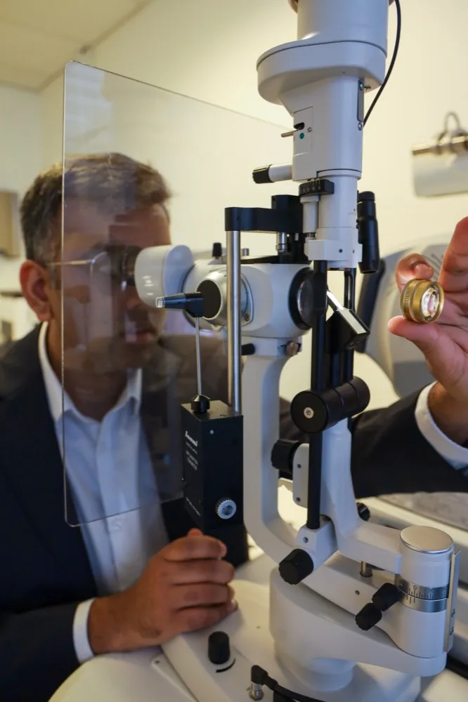
OCT has become an essential exam in ophthalmology. In a matter of seconds, without contact or pain, it produces true cross-sections of the retina and the optic nerve, like a scan of the eye. It makes it possible to detect diseases such as AMD, diabetic macular oedema or glaucoma very early, often before vision even declines. Here is what this exam is used for, how it is performed and why it holds a central place in the follow-up of many eye conditions.
Straight answer: a painless scan of the eye
Key point. OCT, or optical coherence tomography, is an imaging exam that produces microscopic cross-sections of the retina and the optic nerve using a beam of light. It is fast, painless, involves no contact with the eye and no injection. It is used to diagnose and monitor many diseases: AMD, diabetic macular oedema, glaucoma, epiretinal membrane, among others. Its precision makes it possible to spot very early abnormalities and to measure their change over time with great accuracy, which guides treatment decisions.
What is an OCT?
OCT stands for optical coherence tomography. The principle relies on a beam of light that scans the retina: by analysing how this light is reflected by the various layers of tissue, the device reconstructs highly precise cross-sectional images. This gives a detailed representation of the retina, and in particular of the macula, the central area responsible for fine vision, as well as of the optic nerve. The resolution is such that the different retinal layers can be distinguished, on the order of a micrometre, something no conventional fundus examination can reveal.
A recent development, OCT angiography, additionally makes it possible to visualise the retinal vessels without any injection of contrast agent. According to a review published in Progress in Retinal and Eye Research, these non-invasive imaging techniques provide a detailed analysis of the retinal microcirculation and are finding growing applications, including in the monitoring of systemic diseases such as diabetes. OCT has thus established itself as a reference tool, both for diagnosis and for surveillance.
What is an OCT used for?

- AMD: OCT detects oedema, fluid pockets and the neovessels of the wet form, and guides the timing of injections.
- Diabetic macular oedema: it precisely measures the thickness of the macula and how it changes under treatment.
- Glaucoma: it analyses the thickness of the optic nerve fibres, whose thinning is an early sign of the disease.
- Other macular conditions: epiretinal membrane, macular hole or vitreous traction are perfectly visualised in cross-section.
One of the great strengths of OCT is its ability to detect abnormalities before they even affect vision. It is also a remarkable follow-up tool: by comparing exams over time, the ophthalmologist objectively measures whether a disease is progressing, stabilising or responding to treatment. A recent review published in Annals of Translational Medicine highlights that OCT imaging of the retina is now central to the diagnosis and monitoring of the main macular and optic nerve conditions.
How is the exam performed?
The exam is simple and quick. You are seated in front of the device, your chin resting on a chin rest, and you are asked to fixate on a point of light. The device then scans the eye with its beam, without any contact or puff of air. Acquisition lasts only a few seconds per eye. In some cases, dilating the pupil with eye drops is useful to obtain higher-quality images: near vision is then blurred and sensitive to light for a few hours, which may be a reason not to drive right afterwards. Apart from this possible dilation, OCT requires neither injection nor any special preparation, and causes no pain.
FAQ
Frequently asked questions
Is an OCT painful?
No. The exam is carried out without contact with the eye, without a puff of air and without any injection. You simply fixate on a point of light for a few seconds. It is a completely painless exam.
Does the pupil need to be dilated for an OCT?
Not always. Many OCT scans are done without dilation. When it is needed for image quality, eye drops are instilled: near vision stays blurred and sensitive to light for a few hours, so it is better not to drive right afterwards.
What is the difference from a conventional fundus exam?
A fundus exam shows the surface of the retina, whereas an OCT produces cross-sections in depth, layer by layer. The two exams are complementary: OCT provides a precision and a quantitative follow-up that direct observation does not allow.
How often should an OCT be repeated?
It depends on the condition being monitored. For wet AMD or diabetic oedema under treatment, it may be repeated often. For simple surveillance, it is more widely spaced. It is the ophthalmologist who adapts the schedule to your situation.
Can OCT help detect glaucoma?
It contributes decisively. OCT measures the thickness of the optic nerve fibres, whose thinning is an early sign of the disease, at a resolution of the order of a micrometre that no conventional fundus examination can reach. It does not replace the clinical examination, but it provides an objective measurement that can be compared from one visit to the next. It is precisely that comparison over time which shows whether the damage is progressing, holding steady or responding to treatment.
What is OCT angiography?
It is a recent development of OCT that shows the vessels of the retina without any injection of dye. Where standard OCT produces cross-sections of the different retinal layers, OCT angiography gives a detailed analysis of the microcirculation. Its applications keep growing, including in the follow-up of systemic conditions such as diabetes. This is one of the reasons OCT has established itself as a reference tool, for diagnosis as much as for monitoring.
Can OCT pick up a problem before vision declines?
Yes, and that is one of its real strengths. Its precision allows abnormalities to be spotted before they affect vision at all: small pockets of fluid in AMD, thickening of the macula in a patient with diabetes, thinning of the optic nerve fibres in glaucoma. It is also a remarkable monitoring tool: by comparing successive scans, your ophthalmologist can measure objectively whether a disease is progressing, holding steady or responding to treatment.
Scientific sources
- Chua J, Tan B, Wong D, et al. Optical coherence tomography angiography of the retina and choroid in systemic diseases. Prog Retin Eye Res. 2024;103:101292. PMID 39218142.
- Langlo CS, Amin A, Park SS. Optical coherence tomography retinal imaging: narrative review of technological advancements and clinical applications. Ann Transl Med. 2025;13(2):17. PMID 40438521.
- Enaholo ES, Musa MJ, Zeppieri M. Optical Coherence Tomography. StatPearls. PMID 39163419
- Le PH, Kaur K, Patel BC. Optical Coherence Tomography Angiography. StatPearls. PMID 33085382
Further reading
- Dry AMD: surveillance and follow-up
- Epiretinal membrane: symptoms and diagnosis
- Diabetes and the retina: follow-up in Paris 13
- Retinal detachment: recognising the emergency
OCT and retinal examination at the Cachan and Paris 13 practices · Tel. +33 1 45 47 08 11
Getting a retinal OCT
If your ophthalmologist has prescribed an OCT, or if you would like an assessment of the retina and optic nerve, book an appointment with Dr Moïse Tourabaly. The exam, fast and painless, is performed at both practices, in Cachan and Paris 13, and complements the clinical examination for the diagnosis and follow-up of retinal diseases and glaucoma.
This article is intended for information and educational purposes. It does not replace a medical consultation. The indication for and interpretation of an OCT are the responsibility of your ophthalmologist. Sources: articles indexed on PubMed.
Written and reviewed by Dr Moïse Tourabaly, ophthalmic refractive surgeon — former chef de clinique (Quinze-Vingts National Eye Hospital).
Last updated: October 17, 2026